Panorex
Full structure, indepth imaging technology.
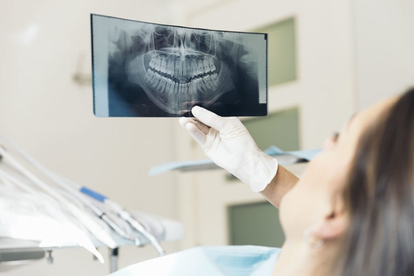
A panorex is a very impressive piece of imaging machinery in that it is capable of identifying many issues and structures that a normal x-ray is not. Initially you will sit in a chair with your chin on a small ledge. Once positioned in the machine, it will rotate around your entire head taking a full 360 degree view of the teeth, head, sinuses and bones.
The ability to view the full structure of your head as a whole is very informative to the dentist. It will allow us to see any potential problems and make sure that everything is functioning as it should be. The panorex is capable of viewing specific types of structural problems, infections or asymmetry among many others.

Intraoral Camera
Real time visual of the inside of your mouth—on the spot.
The intraoral camera is an amazing diagnostic tool for viewing different angles in the mouth. The technology gives us the ability to view the entire mouth on a monitor (video), together, so that we can get a closer look at any potential issues or problems that may arise. These digital images are also excellent for gaining procedure acceptance from insurance companies.
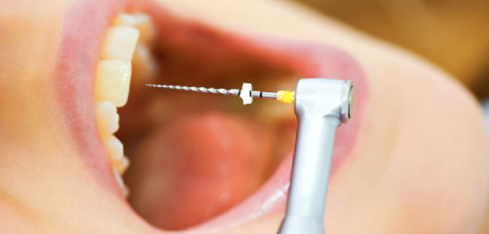
Rotary Endodontics
Easier and often faster root canal process.
Rotary Endodontics is a way of performing the root canal utilizing a specific electrical hand-piece. This tool often makes the process faster and allows the dentist to perform the process with greater ease.
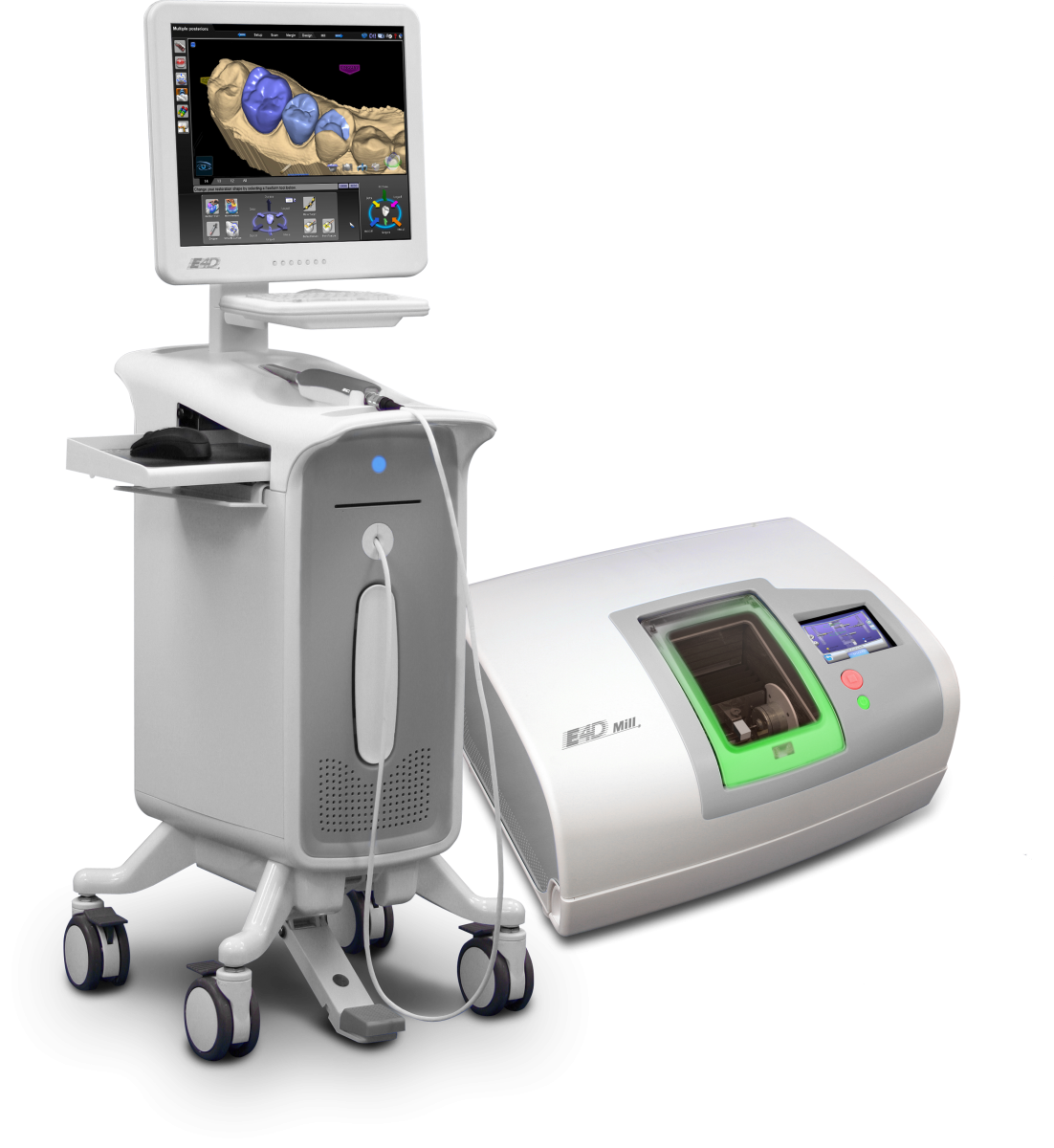
CEREC
Restoration in an hour, start to finish!
CEREC is an advanced dental technology that is utilized for the restoration of decayed, cracked, or chipped teeth. CEREC can create full crowns, inlays, onlays, and veneers. The CEREC machine crafts a restoration in a matter of minutes. CEREC restorations are made of compressed porcelain.
- One visit restoration process.
- No uncomfortable impression material to bite into.
- No temporaries to wear.
Procedure
The procedure for placement of a CEREC restoration is very simple.
- Remove all decay from the tooth.
- Shape the tooth in preparation to take a digital picture.
- The tooth is then sprayed with a very fine powder. This allows the digital camera to take an acceptable image. Once this image is captured, the tooth will appear on a computer screen in 3D. This will allow the doctor to design the restoration right in front of you.
- Once the design is completed, the CEREC will mill the restoration. This step takes approximately 15 minutes. You can actually watch this process if you would like.
- When the restoration is finished milling, the doctor will place the restoration.
The entire process should take about an hour.
Oral Cancer Screenings
Modern technology can pinpoint potential problems early.
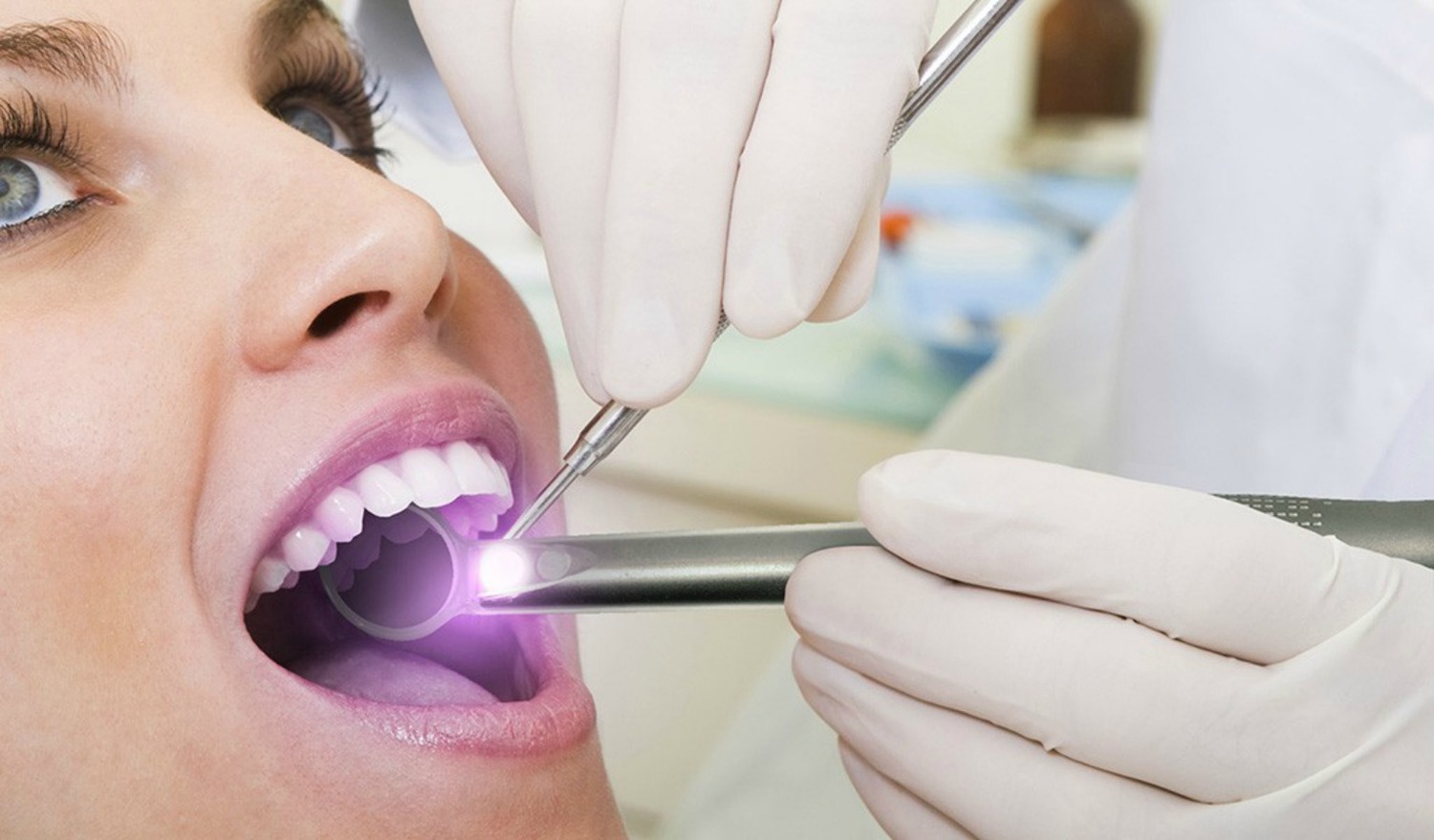
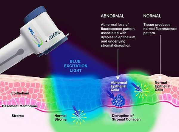
Oral cancer screenings are a very important part of the dental visit for a patient. With the advances in modern technology, we are now able to pinpoint the start of a potential problem much earlier in its evolution. This ability to do so is extremely important in being able to treat any issues prior to them becoming a major irreversible problem.
The oral cancer screening is often completed with an ultraviolet light or similar device that allows us to view issues that can’t always be detected by the human eye under normal conditions.
Certain lifestyle choices can have a great impact on the health of tissues and your overall health in the mouth. If you are a smoker or heavy drinker, make sure to get regular screenings when you visit the dentist.

Digital X-Rays
An accurate, closer look with less radiation.
A digital x-ray allows the dentist to take an image of the tooth or teeth and put it into an imaging program. Within this imaging program, there are a number of tools that will allow the dentist to take a very close look at the teeth and surrounding structures with amazing accuracy.
As a benefit to the patient, the digital x-ray also provides nearly 80% less radiation than a standard x-ray. A digital version of the x-ray is much more sensitive to this radiation and has been specifically designed with the patient in mind.
Magnetic Mallet
Dental surgery and implantology made easier and less painful.
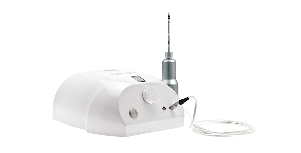
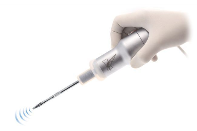
Designed to be used in dentistry surgery and implantology, the Magnetic Mallet gives our dentist a series of options for all advanced bone augmentation procedures such as sinus lift, split crest and ridge expansion, and avoids the use of milling and drills.
It comes with a range of specific instruments that simplify the extraction of roots, implants and impacted teeth, enabling the alveolar integrity preservation.
With the Magnetic Mallet, we can operate with greater visibility and control, preserving the bone and ensuring the greatest possible comfort for the patient both in complex implant surgeries and simple extractions.
This is why Magnetic Mallet innovative, safety and ergonomic features make it such a versatile device, enabling the majority of surgical practices to be performed more easily, safely and quickly while still achieving satisfying and expected results.
Advantages for the patient
- No more BPPV disorder
- Less trauma during surgery
- Faster recovery
- No bone loss
- Poor bone quality is improved
Advantages for the surgeon
- Better visibility and control
- Simplified bone augmentation procedures
- Precision positioning and alignment of osteotomes
- Higher initial implant stability
- Limitation of bone milling
- More implant sites
- No need to perform sinus lifts with Caldwell-Luc
- Alveolar integrity preservation
- Better access to the posterior maxilla
- Simplified lower jaw surgery
- Faster techniques
- Assisted implant insertion
Cone Beam
The best three-dimensional image from teeth to tissue.
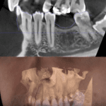
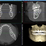
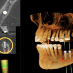
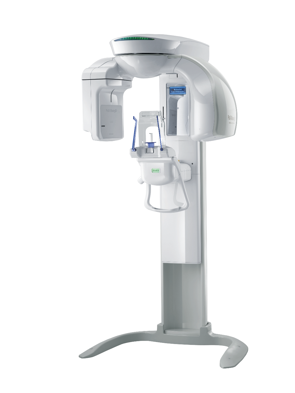
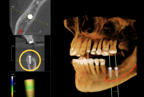
Our new, Dental cone beam computed tomography (CT) is a special type of x-ray device. It is used when regular dental or facial x-rays are not sufficient. This latest technology produces three dimensional (3-D) images of your teeth, soft tissues, nerve pathways and bone in a single scan.
Dental Cone Beam CT is commonly used for treatment planning of orthodontics and dental implants. Cone Beam CT Imaging is also useful for more complex cases that involve:
- Surgical planning for impacted teeth.
- Diagnosing temporomandibular joint disorder (TMJ).
- Evaluation of the jaw, sinuses, nerve canals and nasal cavity.
- Detecting, measuring and treating jaw tumors.
- Determining bone structure and tooth orientation.
- Locating the origin of pain or pathology.
- Cephalometric analysis.
- Reconstructive surgery.
- Accurate and safe placement of dental implants.

